At the fibular neck, the common peroneal nerve lies close to the bone. Here it is at high risk of injury from direct compression.

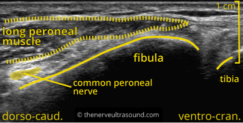
Distal to the fibular neck the common peroneal nerve divides into the deep peroneal nerve and the super- ficial peroneal nerve.
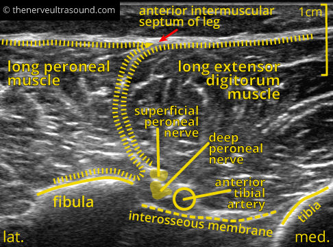
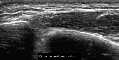
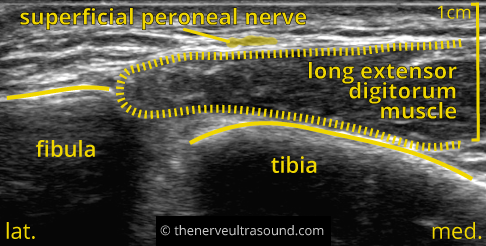
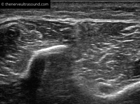
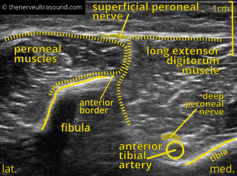
In the distal lower leg, the superficial peroneal nerve runs in the septum between the peroneal muscles and the extensor digitorum longus muscle to reach a superficial position. To find the superficial peroneus nerve at this position look for the anterior border of the fibula bone “pointing” with the intermuscular septum to the nerve at its superficial posion.
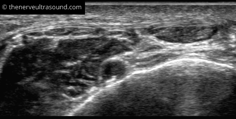
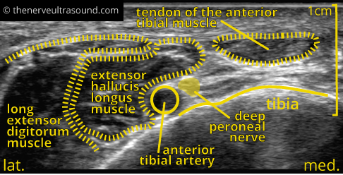
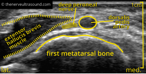
In the same section the deep peroneal nerve may be seen in the depth near the anterior tibial artery.
The superficial peroneal nerve finally pierces the fascia to run subcutaneously. It subsequently divides into the medial dorsal cutaneous nerve and the intermediate dorsal cutaneous nerve.